The Complete Guide to Drop Foot: Understanding Causes, Navigating Treatment, and Living Well
Drop foot is more than an inconvenience—it fundamentally changes how you move through the world. Every step requires conscious effort, every curb becomes an obstacle, and the fear of tripping can make you hesitant to do the activities you once enjoyed without a second thought.
If you or someone you care about is living with drop foot, understanding the condition is the first step toward reclaiming mobility and confidence. This comprehensive guide will walk you through everything from the underlying causes to the latest treatment options, helping you make informed decisions about your care.
What Is Drop Foot?
Drop foot—also called foot drop—is not a disease itself but rather a symptom of an underlying neurological or muscular problem. It describes the inability to lift the front part of your foot, specifically the action called dorsiflexion where you pull your toes upward toward your shin.
This seemingly simple movement is crucial for walking normally. During the swing phase of your gait, dorsiflexion keeps your toes from dragging on the ground. When this ability is compromised, the foot hangs limp or drops down, forcing people to compensate by lifting their knee higher than normal in what clinicians call a "steppage gait."
The condition can affect one foot or both, and it may be temporary or permanent depending on the underlying cause. Some people experience gradual onset, while others develop drop foot suddenly after an injury or surgery.
Recognizing the Symptoms
The hallmark signs of drop foot include foot dragging or scraping along the ground while walking, an exaggerated stepping pattern where the hip and knee lift higher than normal, a slapping sound when the foot hits the floor, and numbness or tingling on the top of the foot or along the outer shin. Many people first notice they're catching their toes on carpet edges, stairs, or uneven surfaces more frequently than before.
The Anatomy Behind the Condition
Understanding why drop foot occurs requires knowing a bit about the nerve and muscle anatomy of your lower leg. The ability to lift your foot depends on three key muscles working together: the tibialis anterior (the primary dorsiflexor), the extensor digitorum longus, and the extensor hallucis longus.
All three muscles are controlled by a single nerve—the common peroneal nerve. This nerve is a branch of the sciatic nerve that wraps around the head of the fibula, the smaller bone on the outside of your lower leg just below the knee. This location makes the peroneal nerve uniquely vulnerable to injury because it lies close to the surface with minimal protective tissue around it.
When this nerve is damaged, compressed, or otherwise compromised, the signals that tell your dorsiflexor muscles to contract are disrupted. The muscles themselves may be perfectly healthy, but without proper nerve input, they cannot function.
What Causes Drop Foot?
The cause of drop foot can originate anywhere along the nerve pathway from the brain to the muscles of the lower leg. A thorough diagnostic workup is essential because treatment depends heavily on identifying where and why the nerve signal is being disrupted.
Peripheral Nerve Pathology
Peripheral nerve compression or injury represents the most common origin of drop foot. Research involving over 1,000 patients found that the vast majority of cases stem from problems at the nerve level rather than central neurological conditions.
L5 Radiculopathy accounts for a significant portion of cases, with studies showing 93.3% of drop foot cases caused by lumbar degenerative disease involve compression of the L5 nerve root. This typically occurs from herniated discs or spinal stenosis in the lower back—conditions that may or may not cause significant back pain.
Direct Peroneal Nerve Injury can occur from compression or trauma at the fibular head. Common culprits include tight casts, prolonged leg crossing, positioning during lengthy surgeries, and direct trauma to the outside of the knee.
Sciatic Nerve Damage higher up in the buttock or thigh can affect the fibers that eventually become the peroneal nerve, causing drop foot along with other symptoms.
Central Neurological Conditions
When the problem originates in the brain or spinal cord rather than peripheral nerves, drop foot presents as part of a larger neurological picture.
Stroke damages motor neurons in the brain, and drop foot affects 20-30% of stroke survivors. The condition typically appears on the side opposite to where the stroke occurred in the brain.
Multiple Sclerosis frequently causes walking difficulties, with drop foot being one of the most common gait problems in MS patients. The demyelination characteristic of MS disrupts nerve signal transmission throughout the central nervous system.
Charcot-Marie-Tooth Disease is an inherited neuropathy that progressively weakens the muscles of the feet and legs. Studies show 100% of patients with CMT develop some degree of foot drop over time, making it a near-universal feature of the condition.
ALS (Amyotrophic Lateral Sclerosis) affects motor neurons, and research indicates 77.2% of patients with lower-limb onset ALS experience drop foot as an early symptom.
Surgical and Traumatic Causes
Drop foot can emerge as a complication following various surgical procedures. Back surgery accounts for 13.9% of cases in some studies, with new neurological deficits occurring in 8.8% of lumbar disc surgeries. Hip replacement carries a 7.8% risk, while knee and foot surgeries contribute 5.1% of cases. Traumatic fractures and accidents account for 11% and 7.1% of cases respectively.
Getting an Accurate Diagnosis
Pinpointing the exact cause and location of nerve involvement is critical because it directly influences treatment decisions and helps set realistic expectations for recovery. A comprehensive diagnostic workup typically involves multiple components.
Clinical Evaluation
Your physician will begin with a thorough physical examination, assessing muscle strength through manual testing of dorsiflexion power. They will evaluate your gait pattern, looking for the characteristic steppage gait or foot slap. Reflex testing and sensory examination help differentiate between different levels of nerve involvement. The distribution of weakness and numbness provides important clues about whether the problem is at the peroneal nerve, L5 nerve root, or higher in the nervous system.
Electrodiagnostic Testing
EMG (electromyography) and nerve conduction studies are essential tools for localizing the lesion. These tests measure electrical activity in muscles and the speed at which nerves conduct signals. They can distinguish between a peroneal nerve injury at the knee versus an L5 radiculopathy in the spine—two conditions that look similar clinically but require different treatment approaches. Electrodiagnostic studies also help determine severity, differentiating between neurapraxia (nerve bruising with good prognosis) and axonotmesis (actual nerve damage requiring longer recovery).
Imaging Studies
MRI scans of the lumbar spine or brain help identify structural causes such as herniated discs, spinal stenosis, tumors, or evidence of stroke. In some cases, ultrasound or MRI of the peripheral nerve itself can identify compression points or masses.
Blood tests may be ordered to rule out systemic conditions that affect nerves, including diabetes, vitamin deficiencies, and inflammatory conditions.
Treatment Options: Finding What Works for You
Management of drop foot is tailored to the underlying cause, the severity of weakness, and the potential for recovery. The therapeutic landscape includes non-invasive orthotics, neurostimulation devices, rehabilitative therapy, and surgical options for appropriate candidates.
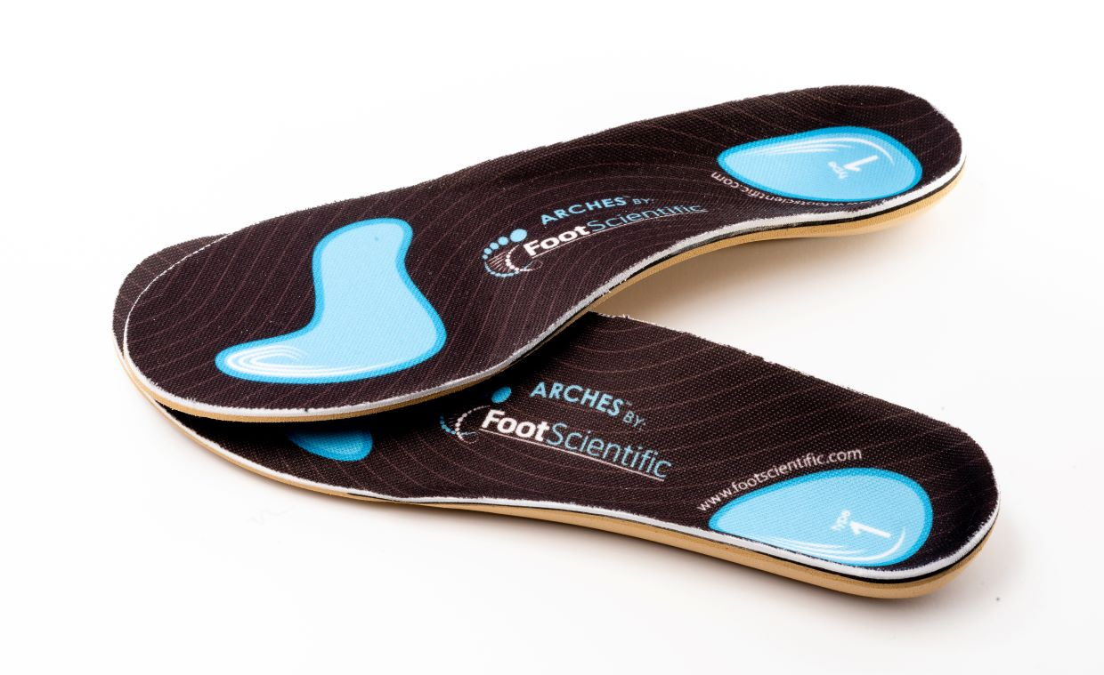
Ankle-Foot Orthoses (AFOs)
AFOs are braces that provide external mechanical support to hold the foot in a neutral position, preventing toe drag during walking. They remain the most common non-surgical treatment for drop foot.
Solid (Rigid) AFOs offer maximum support and completely immobilize the ankle. They are typically prescribed for severe weakness or significant spasticity where any ankle motion would be problematic.
Articulated (Hinged) AFOs allow for controlled, limited ankle motion. They work well for patients who retain some muscle function and need less restrictive support.
Carbon Fiber AFOs provide a lightweight option with energy-return properties. Studies show they can lower energy expenditure by 15% compared to walking without a brace, making them attractive for more active patients.
The Compliance Challenge
Despite their effectiveness when worn, traditional AFOs face a significant limitation: many patients struggle to use them consistently. Research shows approximately 37% of patients demonstrate poor adherence to prescribed AFO wear.
The reasons are practical and understandable. Heat and sweating is reported as a major comfort issue by 87% of AFO users. The braces require larger, often specialized footwear that many patients find unappealing. Bulk and visibility can cause self-consciousness, while some designs create pressure sores. The difficulty of putting on and taking off certain AFOs independently poses challenges, particularly for patients with limited hand function or flexibility.
This non-compliance negates the brace's benefits. A patient who leaves their AFO in the closet because it's uncomfortable or doesn't fit their shoes still faces the same fall risk and mobility limitations as someone without any brace at all.
Modern Alternatives to Traditional AFOs
Recognizing the limitations of traditional AFOs, newer orthotic solutions have emerged that address the primary complaints driving non-compliance.
Functional Electrical Stimulation (FES) uses a device to send electrical impulses to the peroneal nerve, actively stimulating the muscles to lift the foot at the appropriate time during walking. FES has shown equivalent functional improvements to AFOs in stroke patients and works best for individuals with intact peripheral nerve pathways.
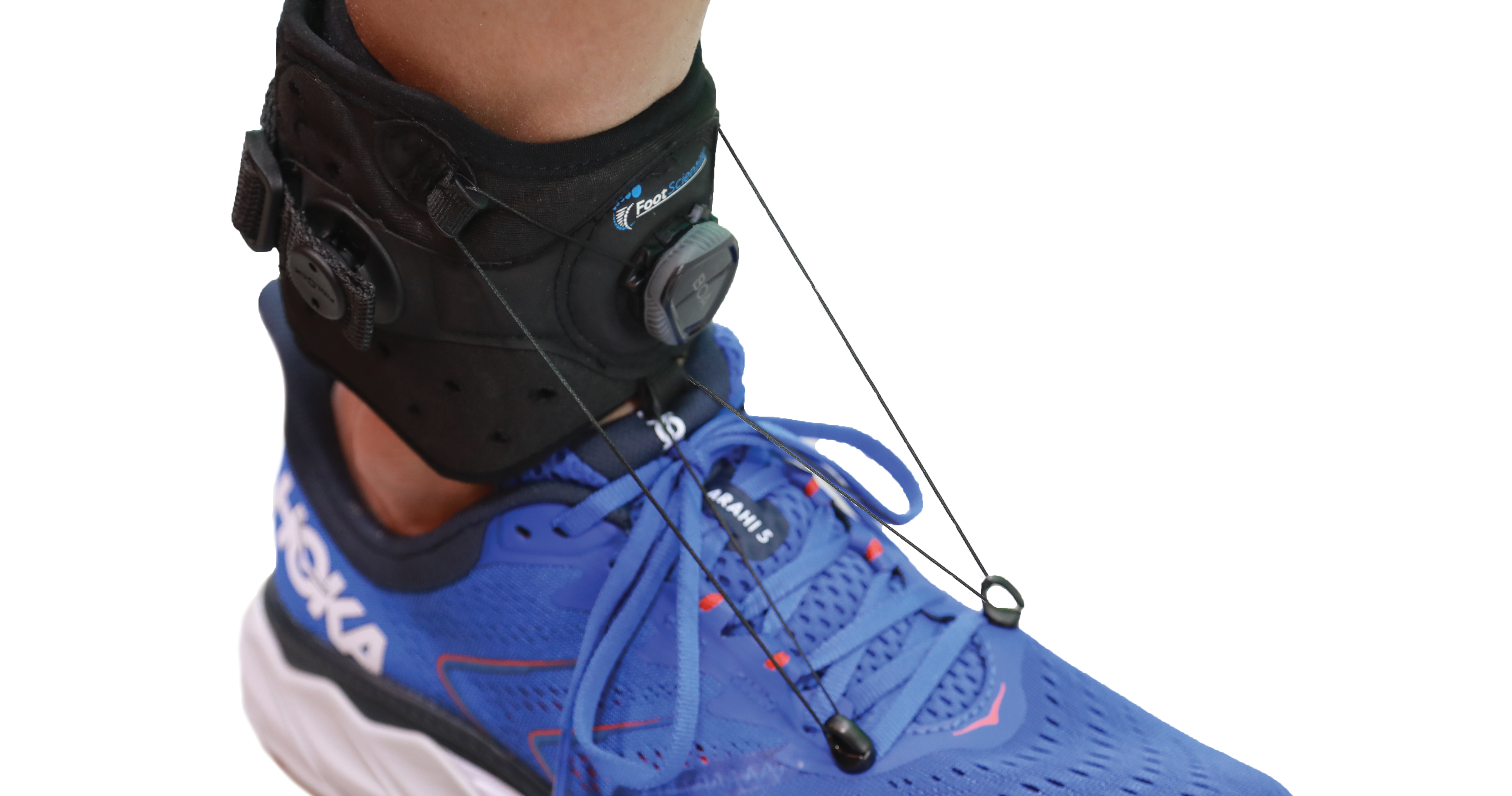
External Drop Foot Braces represent a fundamentally different design philosophy. Rather than fitting inside the shoe, these braces attach to the outside of existing footwear. This approach eliminates the in-shoe bulk and heat that drive so many complaints about traditional AFOs. Patients can continue wearing their own sneakers, boots, or even sandals. The BOA® Fit System technology in some designs allows micro-adjustable lift control, letting users tighten or loosen support throughout the day based on activity level or fatigue.
Physical Therapy
Regardless of what orthotic solution is chosen, physical therapy plays an essential role in drop foot management. A skilled therapist can help strengthen the dorsiflexor muscles if any function remains, along with the compensatory muscles that help maintain stability. Gait retraining teaches patients how to walk safely and efficiently with their brace. Range of motion exercises prevent the ankle from developing contractures that would further limit function. Balance training reduces fall risk beyond what bracing alone provides.
Surgical Options
Surgery is considered when a correctable cause is identified or when conservative treatments have failed. Nerve decompression can relieve pressure on a compressed peroneal nerve, potentially restoring function if the nerve itself is not severely damaged. Tendon transfer surgery—most commonly the posterior tibial tendon transfer—reroutes a healthy tendon to restore dorsiflexion when the original muscles are permanently non-functional. Studies show 73-87% of patients achieve good or excellent results with this procedure. Ankle fusion may be considered for severe cases with joint instability.
Understanding Your Prognosis
One of the first questions patients ask is whether they will recover. The honest answer is that it depends heavily on the underlying cause and the severity of nerve damage.
Prognosis by Injury Type
Neurapraxia refers to temporary nerve dysfunction without structural damage—essentially a "bruised" nerve. The prognosis is excellent, with recovery typically occurring within 2-3 months as the nerve regains normal function.
Axonotmesis involves actual damage to the nerve fibers while the outer nerve sheath remains intact. Recovery is slower, occurring over 6-12 months as the nerve gradually regenerates at approximately one inch per month.
Neurotmesis means complete nerve disruption and carries a poor prognosis without surgical repair.
Prognosis by Cause
Post-stroke drop foot sees most functional recovery in the first 3 months after the stroke, though improvement can continue for up to a year. Long-term bracing is often needed to maintain safe mobility.
Post-surgical cases may improve anywhere from 6 weeks to 12 months after the procedure, with better outcomes for patients who had shorter symptom duration before surgery.
Progressive conditions like MS and CMT typically require lifelong management focused on maintaining function rather than expecting recovery, as the underlying disease process continues.
Signs of Improvement
Recovery happens gradually. Positive signs to watch for include increased ability to lift the foot (even small improvements matter), less frequent tripping or catching of toes, a more natural walking pattern with less need to lift the knee excessively, and return of sensation on the top of the foot if numbness was present.
Living Well with Drop Foot
A drop foot diagnosis doesn't mean giving up the activities and independence you value. With proper management and the right tools, most people can maintain active, fulfilling lives.
Footwear Considerations
If you use a traditional AFO, you'll need footwear large enough to accommodate the brace—typically shoes one to two sizes larger than normal with removable insoles. External braces offer more flexibility, working with your existing supportive shoes. Regardless of brace type, look for shoes with firm heel counters that hold the foot securely, lightweight construction that doesn't add unnecessary weight, and good traction to reduce slip risk.
Staying Active Safely
Many activities remain perfectly safe and beneficial with drop foot. Walking with your brace provides cardiovascular exercise and maintains mobility. Stationary cycling eliminates trip hazards while providing excellent leg exercise. Swimming and aquatic therapy take weight off the joints while building strength. Always consult with your healthcare team before starting new activities, and consider working with a physical therapist to identify safe modifications for activities you enjoy.
Driving Considerations
If your right foot is affected, driving may require modifications or evaluation. A professional driving assessment can determine whether adaptive equipment is needed. Many patients with left foot drop foot continue driving their regular vehicle without modification.
Fall Prevention at Home
Simple modifications significantly reduce fall risk. Remove throw rugs and secure carpet edges. Improve lighting, especially in hallways and stairways. Install grab bars in bathrooms. Use night lights for trips to the bathroom. Keep frequently used items within easy reach to avoid stretching or stepping on tiptoes.
Finding a Solution That Fits Your Life
Drop foot presents a daily challenge to mobility, but it is a manageable condition. The goal of treatment extends beyond simply preventing falls—it's about restoring function, maintaining independence, and preserving your confidence to engage with the world.
The most effective solution is one you'll actually use consistently. A technically superior brace that sits in your closet because it's uncomfortable or limits your footwear choices isn't helping you. Modern bracing options, including external designs that work with your existing shoes, have expanded the choices available to patients who struggled with traditional AFO compliance.
Work with your healthcare team to understand your specific diagnosis, explore all available treatment options, and find an approach that fits your lifestyle. Effective management is about finding a solution that fits your life—not forcing your life to fit a solution.
Ready to Explore Your Options?
If you're living with drop foot and looking for a bracing solution that won't limit your footwear choices, the Elevate Drop Foot Brace was designed by an orthopedic surgeon specifically to address the comfort and usability issues that lead to AFO abandonment.
Shop Elevate Drop Foot Braces →
Frequently Asked Questions About Drop Foot
Whether drop foot is permanent depends entirely on the underlying cause. Cases caused by temporary nerve compression (neurapraxia) often recover fully within 2-3 months. Drop foot from nerve damage (axonotmesis) may improve over 6-12 months as the nerve regenerates. However, drop foot caused by progressive neurological conditions like multiple sclerosis or Charcot-Marie-Tooth disease typically requires ongoing management rather than expecting a cure. An accurate diagnosis through electrodiagnostic testing helps your healthcare provider give you realistic expectations for your specific situation.
Traditional AFOs (ankle-foot orthoses) fit inside your shoe, requiring larger footwear and creating issues with heat and bulk. External drop foot braces like the Elevate Brace attach to the outside of your existing shoes, allowing you to wear your own sneakers, boots, or sandals. External braces eliminate the skin contact issues that cause 87% of AFO users to complain about heat and sweating. The BOA® Fit System in modern external braces also provides micro-adjustable lift control that traditional AFOs cannot offer.
If your left foot is affected, most people can continue driving their standard vehicle without modification. If your right foot is affected, driving safety depends on whether you can operate the gas and brake pedals reliably. A professional driving evaluation is recommended to determine if adaptive equipment is needed. Many drop foot patients successfully drive using left-foot accelerator modifications or hand controls. Always consult with your healthcare provider and consider a certified driver rehabilitation specialist evaluation before returning to driving.
Physical therapy exercises for drop foot focus on strengthening the dorsiflexor muscles if any function remains, maintaining ankle range of motion to prevent contractures, and training compensatory muscles for stability. Common exercises include ankle dorsiflexion against resistance, toe raises, heel walks (if able), and balance training. Gait retraining helps you walk more efficiently with your brace. Learn more in our guide to physical therapy exercises for drop foot. Always work with a licensed physical therapist to develop an appropriate exercise program for your specific condition.
Recovery from drop foot happens gradually, and early signs can be subtle. Positive indicators include any increased ability to lift your foot (even small improvements), less frequent tripping or toe-catching, a more natural walking pattern without needing to lift your knee as high, reduced fatigue when walking, and return of sensation on the top of your foot if numbness was present. Keep a simple log of your daily walking experiences to track patterns over weeks rather than expecting day-to-day changes. Your physical therapist can perform objective strength testing to measure progress.